Bone grafting with bone substitute materials and sinus lift
Placing an implant requires sufficient bone volume. A bone deficit can be made up with the patient's own bone.
Bone substitute materials
A smaller bone deficit can be filled with bone substitute material. The defect is filled at the same time as the implant is placed, often with bone chips mixed in, and the operating area is protected with a membrane.
Placing bone substitute material after an extraction can also counter the breakdown of bone. For grafting, the implantologist can harvest bone in the form of blocks or chips from various areas of the mouth – edentulous areas, the angle of the jaw, the chin – and place it in the area to be built up. Biologically, the patient's own bone is the ideal material, but the drawback of this approach is obvious: it creates a second wound with the familiar disadvantages of pain and risk of complications. The aim is therefore to use a material as close as possible to the body's own bone that does not require a second procedure. Bone substitute materials of various origins are available.
bovine (animal),
synthetic,
synthetic combined with porcine collagen (animal)
All bone substitute materials closely resemble the body's own bone in structure and/or composition. They serve as a scaffold for the blood vessels and cells that matter for bone regeneration and new bone formation. After placement, newly formed bone gradually grows into the substitute material, which is thereby integrated into or remodelled as the patient's own bone.
How long this takes depends on a great many factors and is complete after roughly six to twelve months. The production of bone substitute materials is subject to strict quality and safety controls, and all such materials must meet national and international safety standards. Your dentist will explain the advantages and disadvantages of the alternative materials.
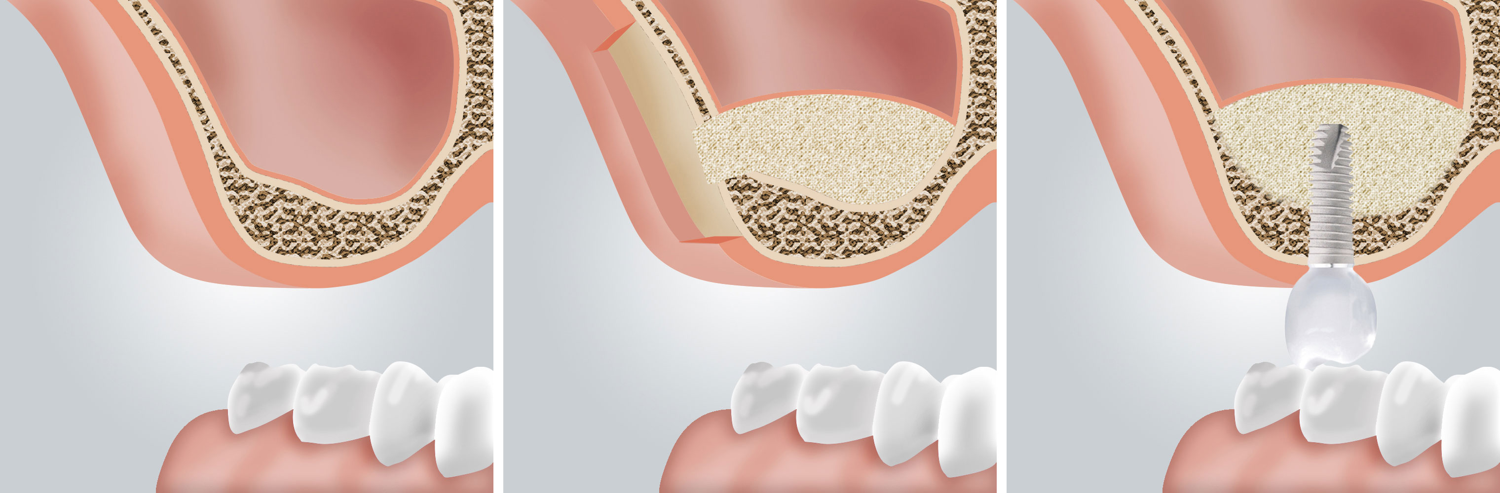
One special form of bone grafting is raising the floor of the maxillary sinus, known as a sinus lift (sinus floor augmentation or elevation). Where there is too little bone in the upper jaw, the sinus can be raised by placing bone or bone substitute material into the maxillary sinus. Where bone is scarce in the posterior upper jaw, a sinus lift is often the only way to create enough bone for implant treatment and a fixed restoration. Where a sinus floor elevation is planned, 3D diagnostics provide detailed information before the procedure about the exact anatomy and about how much bone has to be built up.
Important neighbouring structures, such as the mandibular canal and the bony boundary of the sinus, can be delineated in all three planes.
A distinction is drawn between the internal and the external sinus lift. The external sinus lift is carried out under general anaesthesia. The sinus is opened surgically through the mouth in the region of the cheek so that bone can be placed under direct vision. In the internal sinus lift the implant channel – the hole drilled for the implant – is used to place the bone material. This channel is carefully broken through while sparing the sinus membrane, so that the sinus itself remains unopened, and the bone material is then placed through it. The additional bone makes it possible to place longer implants straight away and to achieve greater stability with them once they have healed.
Bone splitting and bone spreading
Bone spreading and bone splitting are alternatives to the classic bone-grafting measures. These surgical methods are used where the alveolar ridge is very narrow. With this approach the ridge does not have to be built up in a separate procedure before an implant can be placed. The techniques widen a ridge that is too narrow with precision, in order to create a better implant site. Osteotomes, or bone spreaders, prepare the bone gently for placement.
Both methods make it possible to place implants with good prospects of success even after bone has been lost, and in some cases without harvesting bone at all. In selected cases implants can be placed immediately.
Collagen membranes
Collagen membranes have been used as medical devices for many years. Simple collagen sponges can support wound healing or protect the socket after an extraction. Thicker collagen matrices can be used to build up the gum, for example to cover root recessions or soft-tissue defects.
Soft-tissue materials may be made of porcine collagen. Collagens are very stable, fibre-forming proteins that occur widely in the body and make up the main constituent of most connective and supporting tissue. Porcine collagen is very similar to human collagen, which encourages natural tolerance and controlled healing. The soft-tissue material is obtained from natural tissue of pigs intended for the food industry and is cleaned and produced by a multi-stage process. After placement, the body's own tissue grows into the soft-tissue material and it is gradually integrated into the natural tissue structure. After a few months the material has been replaced completely by the patient's own tissue. No further procedure to remove it is needed.
Foundation
When there is not enough jawbone
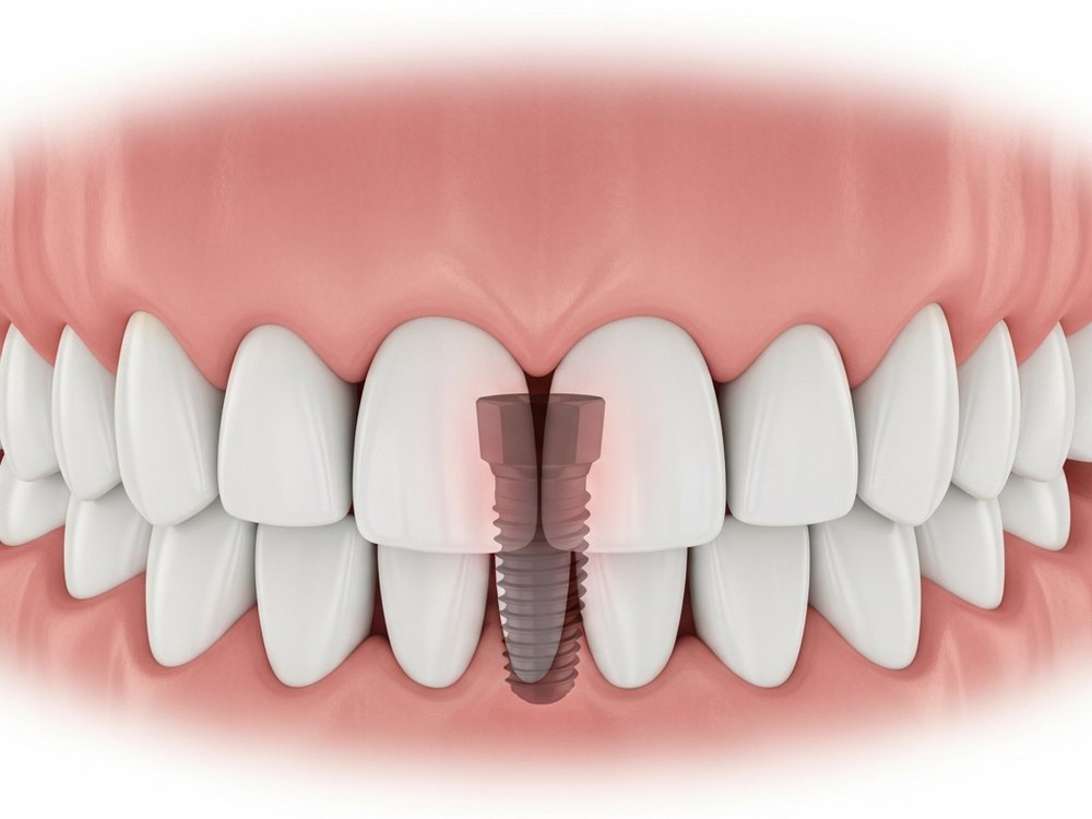
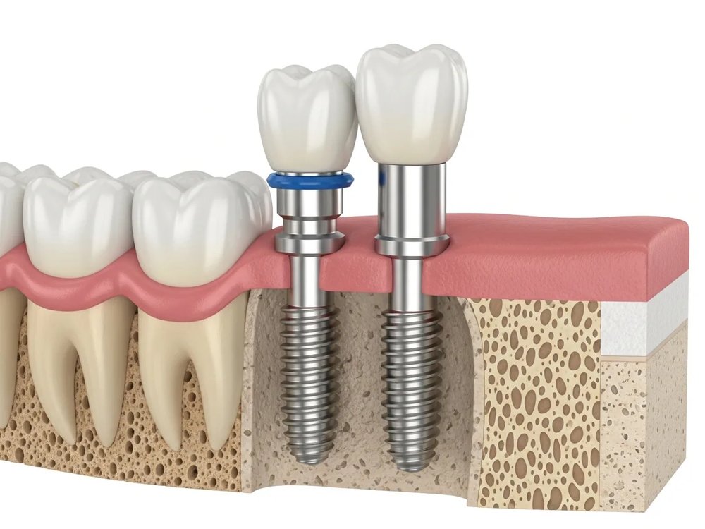
Bone grafting, or augmentation: when teeth are lost, the body breaks down the bone that is no longer loaded. Placing an implant requires sufficient bone volume. Patients who have lost bone after a long period without teeth can be helped too. A bone deficit can be made good in an additional procedure in which the patient's own bone is harvested and grafted. Once healed, this tissue forms a stable foundation for the implants.
The body's own material
The patient's own bone
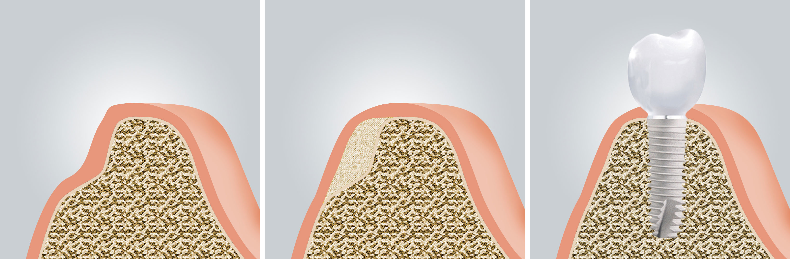
Ridges that are too narrow or too flat, and atrophied areas of the jaw, can be built up with the patient's own bone. The harvested bone is fixed with special screws and generally has to heal for several months. During that time it is covered with a membrane so that stable bone substance can form. Only then are the implants placed. Where the bone deficit is large, grafting uses tissue taken from the patient's own body. Smaller blocks of bone can be harvested from the lower jaw, the chin or the wisdom tooth region and placed directly. Where more bone is needed, it is harvested from the iliac crest in an additional procedure.
The key points at a glance
- When teeth are lost the body breaks down the bone that is no longer loaded; placing an implant requires sufficient bone volume.
- A smaller bone deficit can be filled with bone substitute material at the same time as the implant is placed.
- Where the deficit is larger, the patient's own bone is grafted – from the angle of the jaw, the chin or the wisdom tooth region, and from the iliac crest where a great deal is needed.
- Grafted bone is fixed with screws, covered with a membrane and generally heals for several months before the implant is placed.
Answered briefly