Downloads Video library News FAQs Dental glossary Contact

Implant diagnostics — dental implants and prosthetics

Three-dimensional diagnostics for safe implant treatment.

Thorough diagnostics are the basis for planning and carrying out dental treatment as precisely as possible. New diagnostic techniques allow patients, dentists and surgeons to achieve implant solutions with a high prospect of success. Where it is needed, modern three-dimensional diagnostics today open up reliable diagnostic and therapeutic possibilities in dentistry.

Three-dimensional implant planning on a model of the lower jaw

Before treatment

Planning and preliminary examination

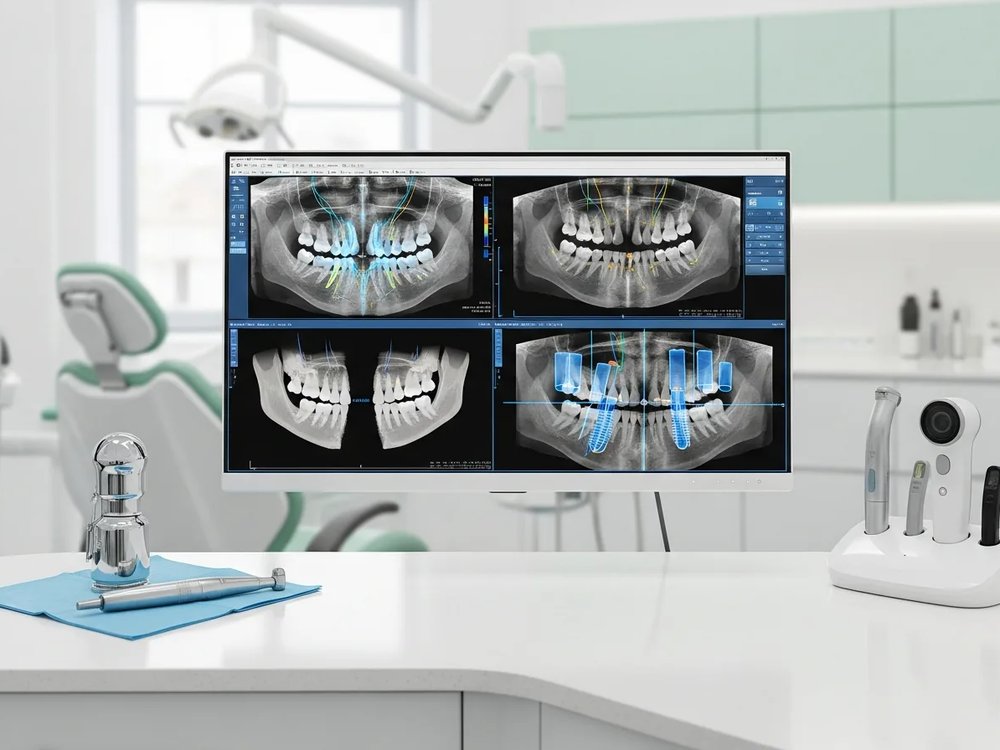
A radiograph gives a first impression of the anatomy, often taken with digital equipment. As far as possible, every potential risk has to be identified before treatment. In implant treatment, diagnostics have a decisive influence on the outcome. Information about the state of the bone at the implant site plays a particularly important part. Is there enough bone, and is its quality sufficient? Bone volume, width and height determine the number of implants. Which implant type, which surface and which length and diameter suit the task? Radiology supplies two-dimensional images with information on the general state of the teeth and the bone.

In more difficult situations these two-dimensional panoramic images are often not enough for reliable implant planning, because the bone can only be assessed to a limited extent. The spatial picture of the bone is missing and the implant site cannot be shown vertically. That carries a risk of injuring anatomical structures such as neighbouring tooth roots, nerves or the maxillary sinus.
The imaging techniques

Which technique is used depends on the question being asked – not every treatment needs a three-dimensional scan.

OPG

The OPG has proved itself in implant diagnostics and in most cases is enough as a basis for planning the implant position. What matters is that the path of the mandibular nerve in the lower jaw is clearly visible and imaged. As a rule the OPG supplies enough information to determine the bone available.

In critical situations – insufficient bone volume, poor bone quality – and for complicated procedures, further detailed diagnostic steps can follow.

More on the OPG

CT

With the development of computed tomography (CT), a further and more precise imaging technique is available. A CT scan is made when a radiograph does not provide the required information about the bone. CT images then make it possible to measure the bone available and to analyse its quality through density measurements. A great advantage are the cross-sectional images a tomogram provides: on these sections the height and width of the bone can be measured directly. The three-dimensional picture of the jaw allows an accurate diagnosis and precise planning of the implant placement.

More on CT

CBCT

Cone beam computed tomography produces cross-sectional images from a series of radiographic exposures. The unit of this special X-ray machine takes these exposures as it travels around the patient's head. They allow a wide range of views on the computer. The three-dimensional, plastic image supports diagnosis, planning and implant placement in critical situations.

More on CBCT

Three-dimensional diagnostics

Three-dimensional imaging of the mouth and jaw region has brought a new quality of care to dentistry. Even in difficult cases, detailed and reliable diagnostics and planning make it possible to carry out more demanding procedures with a promising prognosis.

More on three-dimensional diagnostics

The key points at a glance

  • Before any placement, a radiograph establishes whether there is enough bone and where the nerves and the maxillary sinus run.
  • The panoramic image (OPG) is enough in most cases to plan the number and position of the implants.
  • Only where the two-dimensional image is not enough is a three-dimensional scan added – CBCT or computed tomography.
  • A 3D scan does not belong in every treatment, but in the cases where it genuinely adds knowledge.

Answered briefly

Frequently asked questions

Not in every case. A panoramic radiograph is sufficient for most implant procedures. A three-dimensional scan is indicated where bone is scarce or the situation is difficult.
The OPG is a two-dimensional panoramic image of the jaw and the usual basis for planning. CT and CBCT produce cross-sectional images and therefore a spatial view; CBCT is the technique developed specifically for the jaw region.
Usually 120 to 250 euros including the report. Statutory health insurers do not reimburse CBCT as part of implant planning.
From the radiograph. The panoramic image mainly shows bone height; width and density come from a three-dimensional scan where needed. That determines how many implants are possible.